Catapult Education Product Review
KaVo's DIAGNOcam™ Vision Full HD
KaVo DIAGNOcam: The Next-gen Cavity Diagnosis Tool
To start with, let’s discuss the nature of decay. Our enamel rods are held together with superglue. When we have sugar, the cariogenic bacteria that live in our mouths consume the sugar, release acid as a part of their metabolism, and that acid dissolves the superglue. This is called demineralization.
The reason that we don’t all need fillings even though most of us consume sugar is because a few hours later, the minerals in our saliva come along and “reglue” the enamel rods together, a process called remineralization.
If a person has an imbalance between remineralization and demineralization, which could be due to a variety of factors, they may start to lose some of those enamel rods and develop a cavity.
What are those factors?
There are probably others, but let’s focus on the ones mentioned above. When I look at that list, I think about several potential solutions that don’t involve a drill, such as:
Example Patient Instructions

That’s a lot of options, and while some dentists choose to use only a few of those options or none at all, there is no doubt that options exist between drill and do nothing.
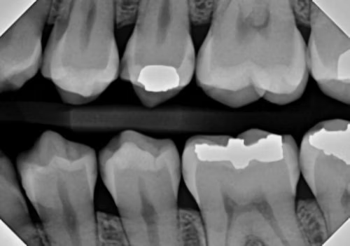
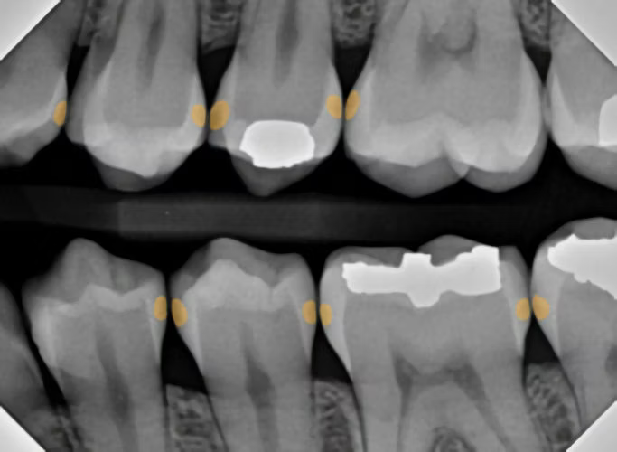
Diagnostic innovation really is necessary to determine whether or not a lesion exists, along with the depth of that lesion. How are these lesions diagnosed? For most dentists, the diagnostic process includes two things: a scaler and an x-ray.
This process, in my opinion, is antiquated. It is difficult to orient a scaler in between teeth, and x-rays in general can sometimes be a nebulous and misleading collection of various shades of gray, highly dependent on the level of thoroughness from the clinician.
I would like to introduce three diagnostic innovations that we probably should be employing, but we may not know about.
The first diagnostic innovation is AI-supported radiographic interpretation
Rather than the discernment of various shades of gray resting on the shoulders of the clinician, who may or may not have adequate time in every different clinical scenario, decay is highlighted by the computer AI software itself, making it a bit more difficult to miss a lesion and highly documenting the degree of enamel or dentin penetration of the lesion.
The second diagnostic innovation is the pH evaluation of cariogenic bacteria inside a lesion
Active decay will have a cavity full of cariogenic bacteria. Consumption of sugar will turn on the metabolic acid production in these bacteria. So, if a person were to chew on a sugary substrate, like a piece of candy, there would be a decrease in pH wherever an active cariogenic lesion exists. More can be learned about this by visiting cavisense.com.
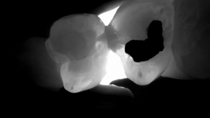
The third diagnostic innovation is transillumination of the interproximal space
Enamel possesses a certain translucency, allowing light to pass through without interruption. A cavity, on the other hand, creates an opacity (a shadow). Therefore, when illumination is applied buccolingually, the cavity is easy to identify. This is valuable for both the identification of dental caries but also for determining the extent of the decay.
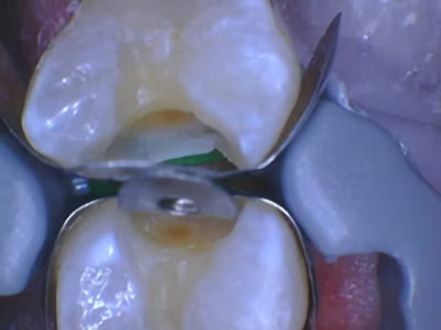
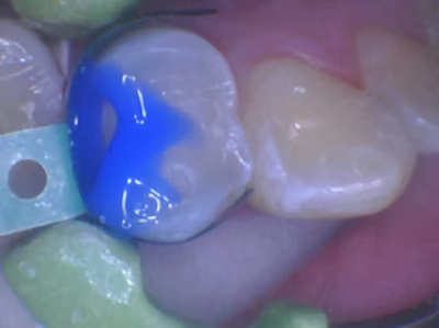
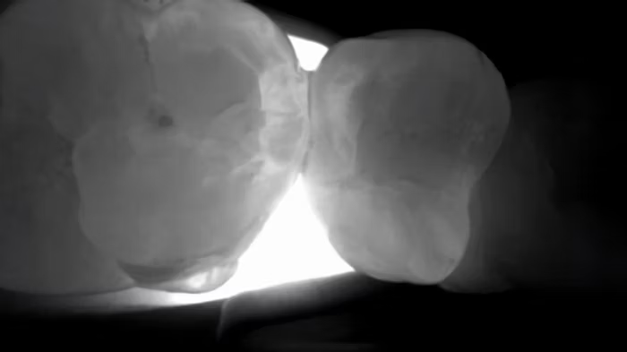
The Catapult Product Evaluation Team, a group of dentists who apply new dental products and innovations into their already active and busy practices, were recently introduced to the DIAGNOcam HD by KaVo. This is a full vision HD camera with three modes. The intraoral mode is similar to most intraoral camera features, but this camera has HD clarity. The two other modes, fluorescence and near-infrared transillumination, are geared more toward diagnosis. The fluorescence mode allows the practitioner to see caries, plaque, and occlusal attrition with greater clarity than with a traditional IO photo. Images in all three modalities can be captured simultaneously and displayed side-by-side.
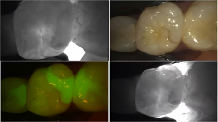
The transillumination mode, however, for me, was the game changer. The DIAGNOCam’s special tip emits near-infrared light at the gingival level, from both the buccal and lingual aspects. The near-infrared light transmits through healthy enamel toward the camera, which has a corresponding bright, white appearance on the resulting image. Areas of carious or demineralized enamel scatter the light, resulting in a corresponding darker region.
Using the same radiograph from above [Figure 4], I transilluminated the questionable areas, yielding highly accurate discernment of the extent and location of various cavities.
The participating Catapult Evaluators were asked the following questions:
The responses to this product review were incredible. Over 85% of survey respondents remarked positively on the questions above. Evaluators were most impressed with the ability of the DIAGNOcam to provide visual confirmation of pathology that simply was not present in the radiographs.
One evaluator remarked, “There was a lingual crack on #3 that was completely missed on the radiograph, and was very difficult to see visually with an intraoral exam. Fluorescence and transillumination made the fracture so obvious. This was helpful both for me and for the patient.”
One evaluator remarked, “I could finally see recurrent decay in three dimensions instead of guessing from the films.” While another evaluator said, “Radiographs pick up the lesion about half the time, and DIAGNOcam showed me enamel changes that I could not see any other way.”


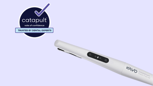
Congratulations: KaVo's DIAGNOcam™ Vision Full HD
Learn More about Ultradent
Products for Your Patients
Say NO to boring CE.
new on-demand and LIVE online courses!